
Menu
Available Technologies
Novel Method for Quantifying Genetic Mutations in Live Cells
Enables quantitative evaluation of cancer-associated genetic mutations based on cell imaging
Background
Cancer arises from genetic mutations, yet even cells with identical genomes can progress differently due to phenotypic variations such as morphology, growth rate, and gene expression. This has increased interest in multi-omics and mechanobiology. However, conventional methods rely on fixed, stained samples, limiting dynamic analysis. Quantitative evaluation of living cells and organoids is therefore essential for advanced and personalized medicine.
Description and Advantages
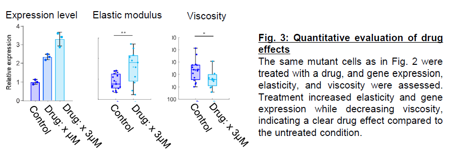
Kyoto University researchers used time-lapse imaging of patient-derived colorectal cancer organoids to analyze deformation during cell division, finding it varies with cancer-related gene mutations (Fig.1). A mathematical model established based on the finding enabled derivation of mechanical properties and quantitative evaluation of these mutations (Fig.2). This technology enables quantitative detection of drug-induced phenotypic changes (Fig.3) and has potential applications in early detection of poor-prognosis mutations, as well as in assessing dose-and irradiation time-dependent responses to inhibitors and radiation.
‣Non-invasive evaluation of living cells
Non-invasive and stain-free, this technology can be applied alongside organoid culture.
‣Analysis based solely on imaging data during cell culture
Quantifying mechanical properties from time-lapse imaging data enables evaluation of
molecular-level alterations.
‣Potential applications in drug efficacy evaluation and personalized medicine
Drug responses in living cells correlate with gene expression levels.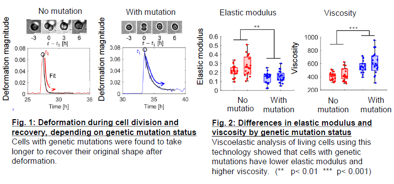
| Offer | ・Patent License ・Option for Patent License ・Collaborative Research |
|---|---|
| Related Links | View PDFView in Japanese |
Have you found what you were looking for?
- Interested in a particular research activity
- Cannot find the information
- Have questions on how to utilize research results
Feel free to contact us and get answers to your questions.
Inquiry- TLO-KYOTO
- Available Technologies
- Novel Method for Quantifying Genetic Mutations in Live Cells

3rd Floor, International Science
Innovation Building, Kyoto University
Yoshidahonmachi, Sakyo-ku, Kyoto
606-8501 JAPAN